Diagnosis & Treatments
How to Diagnose a Bleed in the Brain
Modern research tools and techniques are giving scientists a more detailed understanding of the brain. To diagnose and evaluate an aneurysm, one or more of the following tests or procedures may be performed:
CT Scan
A computerized tomography scan (CT Scan) is a quick tool to visualize the brain tissue with low-dose x-rays. It is a large machine, painless procedure that uses X-rays to generate a 3 dimensional image. A liquid dye that can be seen on an X-ray is injected into an arm vein to outline the aorta or artery on the CT scan. The CT scan images can be used to determine the size and shape of an abdominal aneurysm more accurately than an ultrasound.
TCD
Transcranial doplar (TCD) assesses blood flow within blood vessels of the brain with a small probe placed on the skull.
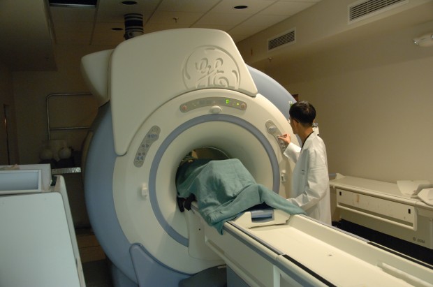
MRI
Magnetic resonance imaging (MRI) gives a precise visualization of brain anatomy to determine the location of the stroke. An MRI uses magnets and radio waves to create images of the inside of the body. It is very accurate in detecting aneurysms and determining their size and exact location.
MRA
Magnetic resonance angiography (MRA) shows the small blood vessels of the brain. Carotid duplex scanning assesses the blood flow within the carotid arteries using sound waves.
PET
A PET scan uses radiation to determine the blood flow within the brain by measuring brain cell metabolism. It provides an image of brain activity.
Cerebral angiography
Angiography also uses a special dye injected into the blood stream to make the insides of arteries show up on X-ray pictures. An angiogram shows the amount of damage and blockage in blood vessels.
Treatment Options
Patients may be referred to a cardiothoracic surgeon, vascular surgeon, or neurosurgeon for diagnosis and treatment of an aneurysm. A cardiothoracic surgeon performs surgery on the heart, lungs, and other organs and structures in the chest, including the aorta. A vascular surgeon performs surgery on the abdominal aorta and on the peripheral arteries. A neurosurgeon performs surgery on the brain, including the arteries in the head, and on the spine and nerves.
A patient's age, overall health, hemorrhage risk and location of the brain aneurysm are considered in determining treatment options. Here are a few of the most common treatment options.
Craniotomy
A craniotomy performed through a surgical opening in the skull. Using an operating microscope and tiny instruments, the surgeon attaches a small metal clip at the base of the aneurysm. Because blood is prevented from flowing into the aneurysm, the chances of it rupturing are greatly reduced. Recovery time typically is four to six weeks.
Endovascular Surgery
Endovascular surgery is performed through a catheter threaded through the blood vessels. Thin platinum wires are pushed into the aneurysm, where they coil into a mesh ball. Because blood clots form around the coils, the chances of it rupturing are greatly reduced. Recovery time typically is two to four days.
Clipping
Clipping is also a common surgical treatment for brain aneurysms. Microvascular clipping cuts off the blood flow to the aneurysm. Once the aneurysm is located, the neurosurgeon uses a microscope to clip the aneurysm’s neck, stopping the blood supply. The clip remains in the patient to prevent future bleeding.
For More Information